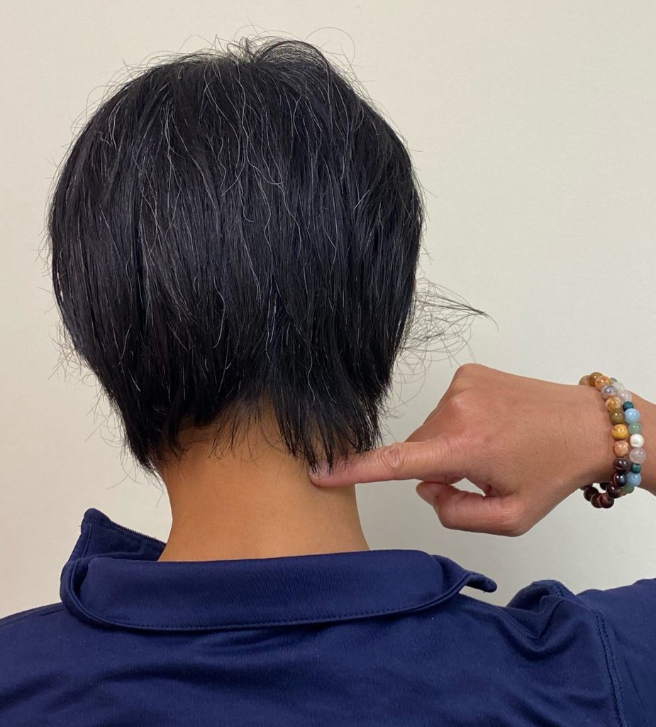
Sometimes neck stiffness, arm weakness, or feelings of pins and needles down your arm can be signs of a “pinched nerve” from the neck.
- Looking over your shoulder while driving
- Gazing up at the birds in the sky
- Sleeping in the “wrong” position
Sometimes, a pinched nerve can even send pain or numbness into the fingers, which makes it feel like an issue throughout your entire arm rather than just the neck. This injury sounds scary, but it can be successfully addressed with conservative treatment and lifestyle adjustment. Physical therapy can definitely help!
Let’s dive into what a pinched nerve means and what causes it.
About Your Neck’s Spine
To understand the cause of a pinched nerve, you need to be familiar with the anatomy of the neck, also known as the cervical spine.
Bony Anatomy
-
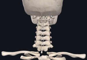
- Cervical Spine | Images courtesy of Complete Anatomy
These joints and their directional orientation allow for the movements of the neck to occur:
- side-bending (ear to shoulder)
- rotation (looking over your shoulder)
- flexion (looking down)
- extension (looking up)
The first two cervical vertebrae are very different from the lower vertebrae. They are known as the atlas (C1) and the axis (C2).
-
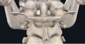
- Atlas & Axis | Images courtesy of Complete Anatomy
These vertebrae are responsible for the majority of the movement of head and neck, assisting us with looking up, down, and over our shoulders.
The lower cervical vertebrae (C3-C7) are fairly similar in shape;
-
- C7 Vertebra (highlighted) | Images courtesy of Complete Anatomy
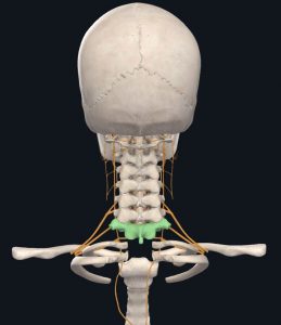
The Nerves
These roots typically referred to as C1-C8 run between the vertebral segments, but the C1 nerve root exits above the C1 vertebrae. This is the only exception.
These nerves, known as peripheral nerves once they exit the spinal canal, are responsible for sending and carrying signals to and from the brain to various parts of the body allowing for proper functioning (think of the skin and muscles).
-
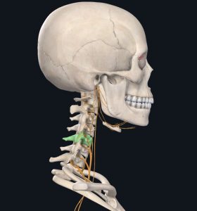
- Peripheral Nerves Leaving the Spine (in yellow) | Images courtesy of Complete Anatomy
If you are otherwise healthy, a disc herniation is often the cause of a pinched nerve.
The vertebral discs allow each segment to easily move on top of one another as well as absorb shock and transmit force through the spinal column to prevent injury. With excessive force or repetitive movements, these discs can sometimes push out of the openings between each vertebrae and encroach on the nerve roots that run through the intervertebral foramen.
However, your body is resilient, and tissues heal over time. A disc has been shown to resorb given the right guidance, which is where physical therapy enters the chat.
Typical Signs and Symptoms of a Pinched Nerve
- A stiff neck or difficulty moving the head & neck
- Weakness in the shoulder, arm, or hand
- Numbness, tingling, burning, shooting, & or throbbing running from the neck down into the shoulder blade, down the arm, and even into the hand and fingers
Each nerve, either motor (supplies input to the muscles) or sensory (supplies input to the skin), has a specific pathway it will follow.
The most common nerve roots affected are C5-C7 due to the fact that as the head bends forward when we look down at our phones, computers, etc. the weight of the head on the neck continues to increase.
There is currently a diagnosis called Text Neck in the medical world that refers to this phenomenon.
According to an article published in the Washington Post, the weight of the head on the neck is normally 10-12 lbs, but with an increase in forward flexion the weight of the head on the neck can increase to 60 lbs!
This added pressure and repetitive tension can result in disc herniation with nerve impingement or arthritic changes.
Other Signs and Symptoms of a Pinched Nerve
Other signs and symptoms that can accompany a pinched nerve include the following and may warrant further follow-up by your primary care doctor:
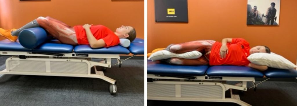
Difficulty sleeping
- Utilizing pillows to support the head and neck and decrease compression of the nerve roots can help. M
ost people wake up during the night to change positions because numbness/tingling usually occurs when pressure is taken off the nerve See below for examples of how to utilize pillows sleeping on your back and sides to support a neutral neck and spine (small blue roll is a cervical roll pictured in the first image below).
Dizziness or lightheadedness and headaches
This is usually caused by tight muscles of the head and neck that frequently stiffen up to protect the injured area. - See our blog on cervicogenic headaches to learn more
In more serious cases, this is caused by blood flow issues to the brain! - If you have a history of cardiovascular disease or difficulty controlling your blood pressure be sure to follow-up with your primary care doctor and notify your PT.
Signs and symptoms that are indicative of serious pathology and require an immediate follow-up with your primary care doctor include:
- Headache that occurs with coughing, physical exertion, or breath holding (valsalva)
- This is usually a sign of a serious condition such as bleeding in the brain (subarachnoid hemorrhage) or a possible lesion/tumor
- Loss of bowel and bladder control or retention of urine or feces
- Weight-loss or gain > 10% within the last month
- Progressive weakness or muscle stiffening that has not improved with conservative care
How to Treat a Pinched Nerve
In severe instances where arthritic changes cause near complete narrowing of the spinal canal or foramen, surgical intervention may be warranted including the following procedures:
- Laminectomy where the lamina, or bony area between the vertebral body and spinous process, and other structures are removed to open up space for the nerve root or
- Discectomy where the herniated disc is removed that is impinging upon the nerve root, sometimes referred to as a ACDF or Anterior Cervical Discectomy and Fusion
- A discectomy is almost always accompanied by a Cervical Fusion to then stabilize the spine by placing a bone graft or implant where the disc was removed.
- Loss of some normal range of motion can occur after a cervical fusion. Unfortunately, 25% of individuals may experience a recurrence of pinched nerve symptoms.
If you are experiencing any of the above signs and symptoms or have been diagnosed with a pinched nerve by your doctor, try physical therapy first. Physical therapy can help you avoid surgery!
If you haven’t received a referral for physical therapy from your doctor, reach out to JACO to see how we can provide you with 1-on-1 care.
Physical Therapy for a Pinched Nerve
At JACO, we individualize your treatment to your specific deficits. An initial evaluation gives us most of the information we need to get you on the right path.
There are many common deficits we see related to a pinched nerve. Many interventions prescribed by your physical therapist could include:
- Improving neck range of motion
- Increasing muscle extensibility via stretching
- Enhancing postural stability by strengthening the deep neck flexors and scapular muscles
- Screening and addressing habits that may contribute to your symptoms
With the combination of our guidance and your program consistency, signs and symptoms can be greatly improved or totally resolved!
See our blog on centralization versus peripheralization to take a closer look at how individualized physical therapy treatment can directly improve radiating numbness, tingling, burning, or throbbing.
Exercise Examples to Treat a Pinched Nerve
If you’re having symptoms, you should get a referral to physical therapy to address your individual needs. However, there are foundational exercises that help most people get a head start.
Until you get to a therapist, try these. If the exercise increases your symptoms, stop and refer to your therapist.
Chin Tuck
The majority of pinched nerves can occur due to excessive load on the lower cervical spine. Performing chin tucks working on strengthening the deep neck flexors in the front of your neck can help alleviate tension on the nerve roots.
-
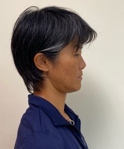
- Chin Tuck – Start Position
-
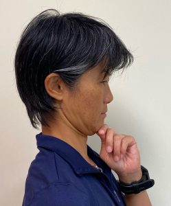
- Chin Tuck – End Position
Steps to perform a chin tuck:
- Lay on your back with your knees bent and neck and shoulders relaxed.
- Gentle tuck your chin towards your throat or think about performing a gentle nod with your chin (think yes and no).
- You should feel a slight stretch at the base of your skull and tightening of the muscles under your chin.
- If you’re having trouble feeling the right muscles working, place two fingers on your chin and slowly push back until you can feel the muscles under your chin tighten up.
- Hold this position for as long as you can up to 30 seconds, or a minimum of 3 seconds.
Repeat 3 sets x10 repetitions.
Nerve Glide
One exercise to decrease the tension on the nerve is to perform a nerve glide.
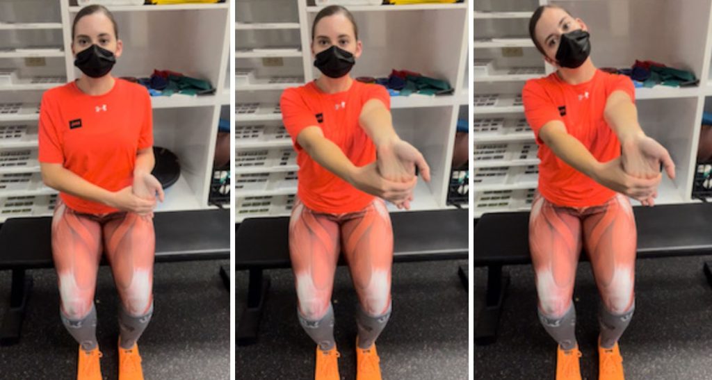
Steps to perform a general nerve glide for the upper extremity:
- Sit in a chair with your back supported with chest proud and shoulder blades gently squeezed together.
- Perform a gentle chin tuck (see exercise above) to decrease stress on the neck.
- Grab your wrist and fingers at your knuckles with your opposite hand and gently pull them back towards you. Stop when you feel a light stretch.
- Straighten your elbow ensuring your shoulder does not elevate. You should feel a gentle stretch along your arm.
- For an increased stretch bring your opposite ear to your opposite shoulder as you straighten your arm.
Perform 3 sets x10 repetitions.
Contact Us Today
Get an evaluation at JACO Rehab at any of our four locations: Honolulu, Mililani, Waikele, or Kapolei.
Written by Jenn Lewis, DPT